Electron Microscopy
Electron microscopy is a specialized field of science used for clinical diagnostic purposes as well as for research. Electron microscopy has been used in all areas of life sciences such as anatomy, biochemistry, botany, cell biology, forensic medicine, microbiology, pathology (especially in renal and tumor pathology), physiology and toxicology. It can achieve an optimum magnification of more than 600,000 times with a resolution of about 0.2nm, thus allowing viewing of fine ultrastructure. The electron microscope (EM) is used not only to visualize biological materials but can also be used to analyse the chemical make-up and physical properties of a specimen.
About Us
The Electron Microscopy Unit provides a centralized microscopy service to all faculties in the University of Malaya as well as to other universities, research institutions and private companies. The Unit is able to advise users on many aspects of electron microscopy.
The EM Unit consists of one electron microscopy room, a darkroom, an ultramicrotomy sectioning area, a processing area and a general laboratory, all housed within six standard modular units of 10’ x 25’ each.
History
The Electron Microscopy Unit (EM Unit) was set up as a central facility, under a vote controlled by the Dean of the Faculty of Medicine in 1969. Previously it was located within two standard modular units of 10’ x 25’ each at the Department of Pathology as it was considered a central location in the Medical Centre. The chairman of the EM committee was the Head of Department of Pathology at that time , Prof. K.Prathap.
The EM Unit was then shifted to a more suitable location within six standard modular units at the post-graduate block in September 2002. It was then placed directly under the administration of the Deputy Dean of Research in October 2004 as it was accorded the status of a central research facility for the Faculty of Medicine.
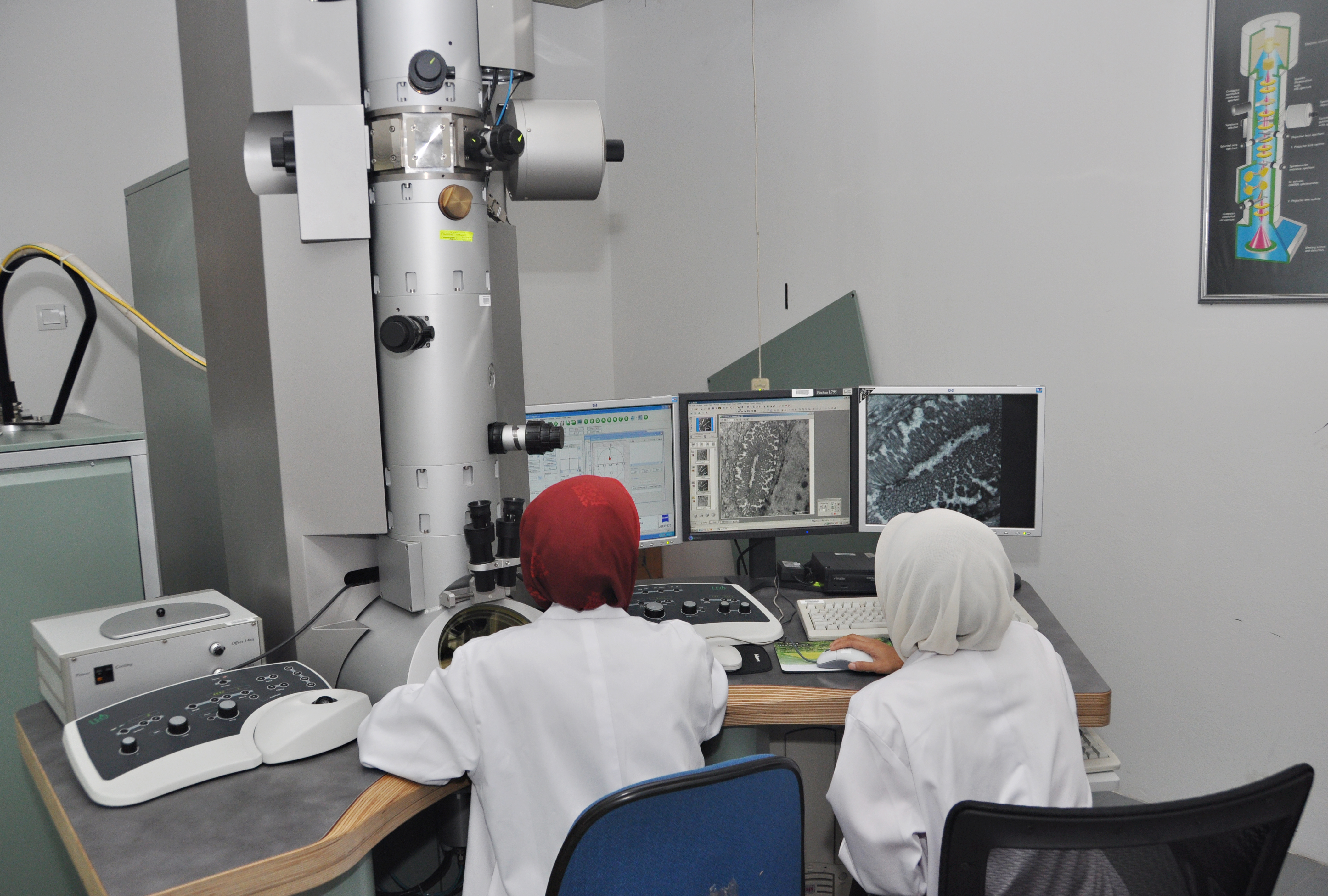
The first Transmission Electron Microscope (TEM) which was the Hitachi HS-8 and other related equipment were bought under a financial grant of RM100,000 from China Medical Board , USA. The Hitachi TEM was boarded out in 1985 and is currently being housed in the Medical Faculty Museum as one of the faculty’s artefacts. The second TEM was the Philips CM-10 which was bought in 1986 and was in use until 2002. The latest TEM is the LEO-Libra 120 purchased in December 2003 at a cost of RM2 million.
Since 1969, the EM Unit has not only contributed towards diagnostic work in the University Malaya Medical Centre (UMMC) and other government and private Hospitals throughout Malaysia but also subserved research endeavours throughout the faculty and University Malaya and other institutions of higher learning throughout the country. Currently, the departments most actively engaged in EM-related research are the departments in the Faculty of Medicine involved in research related to electron microscopy are Departments of Anatomy, Orthosurgery, Pharmacology, Pathology, Parasitology , Microbiology and Oral Microbiology. The departments of Pathology and Microbiology are also actively involved in diagnostic-related electron microscopy.
Team Members
- Head of Unit: Dr Leo Bey Fen
- Deputy Head of Unit: Associate Prof Dr Anwar Norazit
- Medical Lab Technologists & Assistant Science Officers:
- Ms Pang Swee Ling
- Ms Azura Aladdin
- Ms Norizreen Fara
- Mr Hamzah Abd Hamid
- Mr Amirul Asraf
Equipment & Services
Equipment:
1. Transmission Electron Microscopy (TEM) LEO Libra-120
- Ultrasctructure viewing
2. Field Emission Scanning Electron Microscopy (FESEM) Quanta FEG 650
- High/Low vacuum
- High/Low vacuum with EDS analysis
3. Ultramicrotome EM UC6/UC7
- Semithin sectioning
- Ultrathin sectioning
4. Sputter Coater Leica EM SCD005
- Gold coating
5. Automated Critical Point Dryer Leica CPD300
- Critical point drying for biological sample
We provide users variety of services and consultation for both clinical and research purposes. The services provided include:
- Preparation of transmission electron microscopy samples (TEM)
- Preparation of semithin and ultrathin tissue sections
- Material sample and negative staining
- Viewing samples using TEM and image capture (TEM Imaging)
- Preparation of scanning electron micrscopy (SEM) samples (FESEM Imaging)
- Critical point drying
- Mounting
- Gold coating
We also offer EM Laboratory technical training, from sample preparation to usage of the electron microscope. A nominal sum is charged for the training and use of the various instruments and consumables.
